Zeta-potential & Particle size Analyzer ELSZneo NEW
|
|
ELSZneo is the high-end model of the ELSZ series. This instrument measures zeta potential and particle size of both diluted and concentrated solutions, and molecular weight of a polymer. As new features, ELSZneo provides a multi-angle measurement to improve the separability of particle size distribution, particle concentration measurement, microrheology measurement, and gel network analysis.
Are you interested in alliance or distributorship for this product? |
- Product
- Principle
- Specification
- Applications
- Option
Product
- Particle size and zeta potential measurement in a wide concentration range from diluted to concentrated solution (~ 40%)
- Multi-angle measurement to carry out particle size distribution with higher separability
- Zeta potential measurement of solid surface in high salt concentration
- Particle concentration analysis using Static Light Scattering
- Microrheology measurement using Dynamic Light Scattering
- Gel network and gel heterogeneity analysis by measuring intensity of scattered light and diffusion coefficient of sample at multiple points
- Standard flow cell which can be used for two types of measurement (Particle size and zeta potential) without replacing samples
- Wide range of measurement temperature from0to 90℃
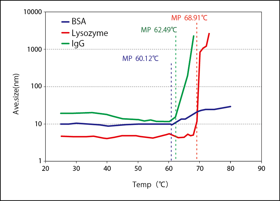
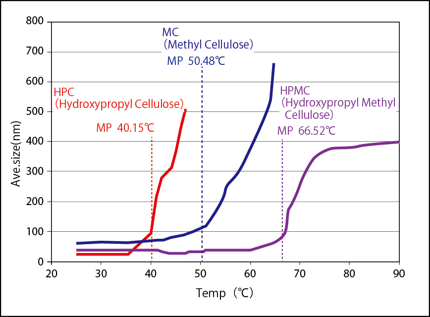
- Temperature gradient function which enables denaturation (melting temperature) analysis for proteins
- Electrophoretic mobility measurement and plot analysis for accurate zeta potential measurement
- Band-pass filter (option)
- Particle Size Distribution
- Zeta Potential
- Molecular Weight
- Microrheology
- Particle Concentration
- Structural Analysis of Gel Networks
| Zeta Potential | No effective limitations | |||
| Mobility | -2×10 -5 ~ 2×10 -5 cm2/V・s | |||
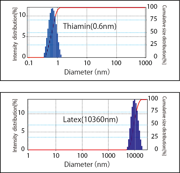
| Particle Size | 0.6 nm ~ 10000 nm*1 (display range:0.1-106nm) |
|||
| Molecular Weight | 340 ~ 2×10⁷ | |||
*1:Minimum value with histogram analysis : 0.2 nm
●Measure conditions
| Temperature | 0 ~ 90℃ | |||
| Concentration | Particle Size : 0.00001 % (0.1ppm) ~ 40 % *2 Zeta Potential : 0.001%~40% |
|||
*2:(Latex112nm: 0.00001 ~ 10%、Taurocholic acid: ~ 40%)
Best suitable for basic and applied research of particle characterization in the field of surface chemical, inorganic, semiconductor, polymer, biotechnology, pharmaceutical and medical as well as surface research of film and flat state sample.
- New functional material
Fuel cell(Carbon nanotube, Fullerene, Function film, Catalyst, Nano-metal)
Bionanotechnology ( Nano capsule, Dendrimer, Drug Delivery System(DDS)), Nanobubble
Biocompatible material
- Ceramic and paints
Ceramic(Silica, Alumina, TiO2)
Surface modification, dispersion control of inorganic sol
Dispersibility, durability and shelf life control of ink, carbon black and organic pigment
Slurry state sample
Color filter
Flocculation research - Semiconductor
Alien objects research on silicon wafer
Interaction research of abrasive/additive on wafer surface
CMP slurry
- Polymer and Chemical
Emulsion dispersion stability to extend product shelf life
Surface modification of latex
Function research of polyelectrolyte
Process control of paper production and Pulp additive research
- Pharmaceutical and food
Emulsion dispersion stability to extend product shelf life
Dispersibility control of liposome and vesicle
Functionality of surfactant(Micelle)
Exosome,
Viruses, Virus-like particles(VLP)
Proteins
Principle
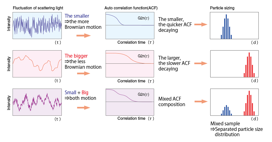
Particulates dispersed in a solution are normally subject to Brownian motion. The motion is slower with larger particles and faster with smaller particles. When laser light illuminates particles under the influence of Brownian motion, scattered light from the particles shows fluctuation corresponding to individual particles. The fluctuation is observed according to the pinhole type photon detection method so that particle size and particle size distributions are calculated.
%20.jpg)
In most cases, colloidal particles possess a positive or negative electrostatic charge. As electrical fields are applied to the particle dispersion, the particles migrate in oppositely charged directions. As particles are irradiated in migration, scattering light causes Doppler shift depending on electrophoretic mobility. This method is called Laser Doppler Method.
%20.jpg)
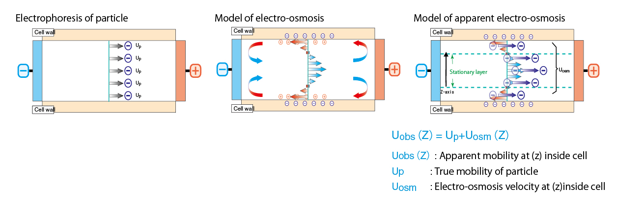
Electro-osmosis is the liquid flow occurred inside cell upon zeta potential measurement. If cell wall is electrically charged, counter ion in medium migrates to cell wall. This is the phenomenon that counter ion migrates to one electrode in the cell wall and to other electrode in the vicinity of cell center. By measuring apparent electrophoresis mobility and analyzing electro osmosis, it becomes possible to obtain correct stationary layer taking stained cell into consideration and to calculate correct zeta potential. (Mori-Okamoto equation)

<Mori-Okamoto equation >
Uobs(z)=AU0(z/b)2+⊿U0(z/b)+(1-A)U0+Up
z:Distance from cell center
Uobs(z):Apparent mobility at (z) inside cell
A=1/[(2/3)-(0.420166/k)]
k=2a and 2b are respectively the horizontal and vertical length of square phase of cell. Here a>b.
Up:True mobility of particle
U0:Average mobility at upper and bottom walls
⊿U0:Gap of mobility at upper and bottom walls
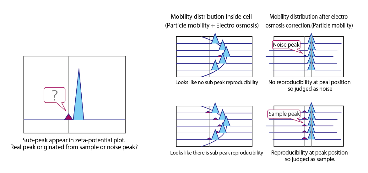
Apparent electro-osmosis measure at multiple points inside cell enables repeatability check of zeta potential and noise or peak determination.
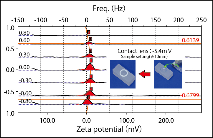
Flat surface cell is configured with box-like quartz cell with flat surface sample attached on it. Measure apparent electrophoresis mobility of monitoring particle at several positions in vertical inside cell and analyze mobility of electro osmosis on solid surface using electro osmosis profile obtained to calculate zeta potential.

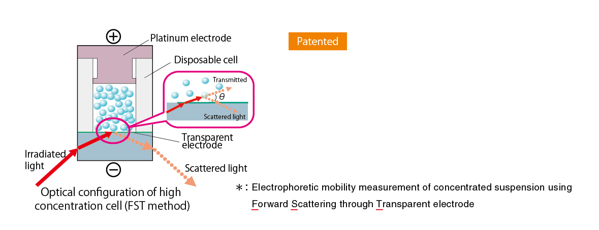
It used to be difficult to measure very condensed or colored sample due to multiple scattering or absorption effect. Currently standard cell of ELSZ series is able to measure wide concentration range of sample. Furthermore, zeta potential of very condensed sample can be measured by FST method*
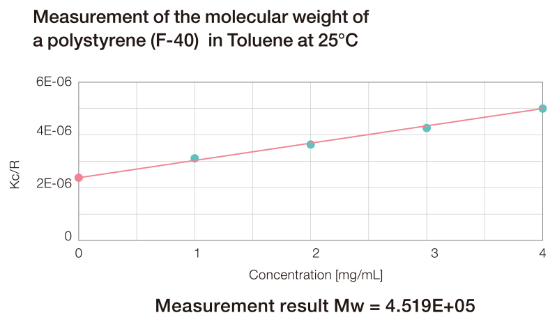
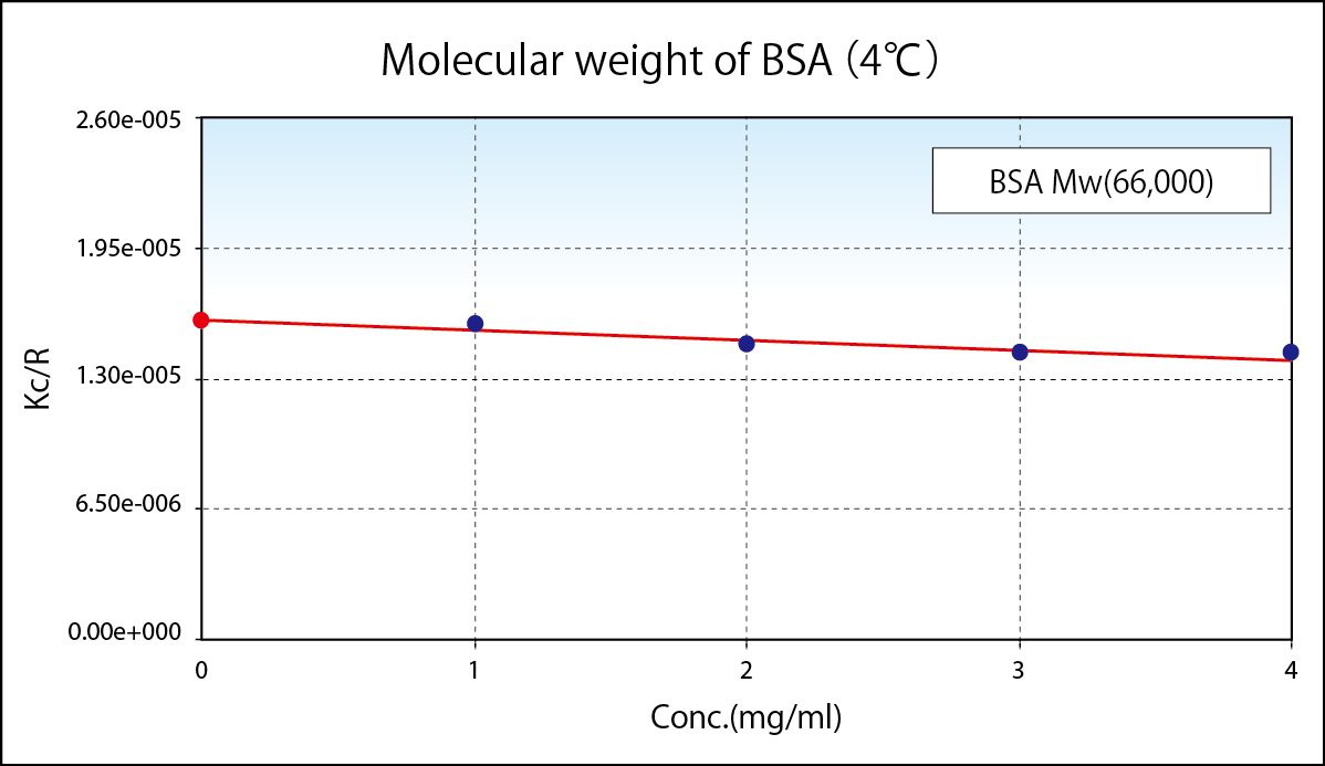
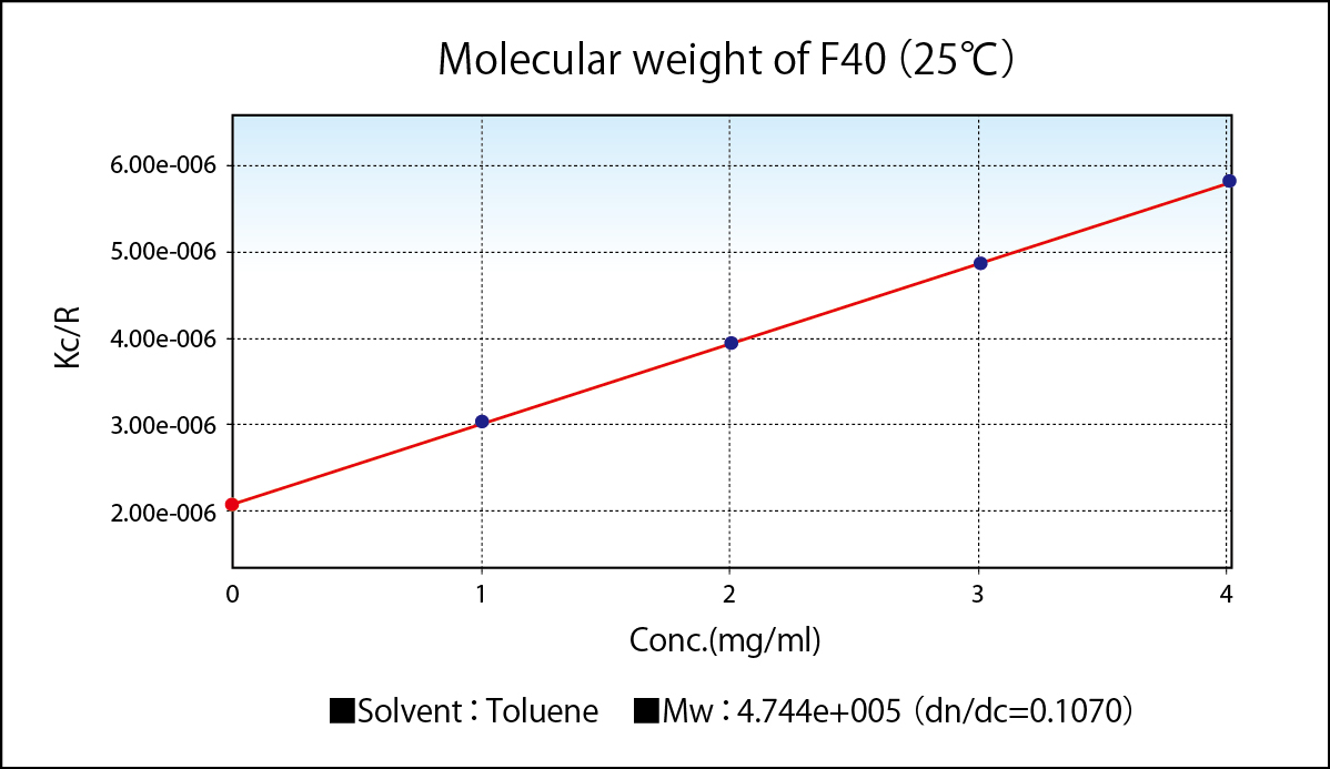
Static Light Scattering Method is renowned as the convenient method to know absolute molecular weight. As a principle, molecular weight is calculated from absolute value of scattered light obtained by irradiated light into colloidal particle. To simplify, from the bigger particle gives the stronger scattering and the smaller particle gives weaker scattering. In reality, scattering intensity to be obtained depends upon concentration, too. So the plotting concentration on horizontal and Kc/R(θ), which is equivalent as a reciprocal number of scattering intensity, on vertical after measuring scattering intensities of variable concentrations is called Debye Plot.
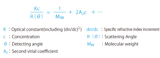
Since angle dependence of scattering intensity appears in the large molecular weight sample, measuring scattering intensity at variable angle(θ) provides more accuracy on molecular weight measurement and radius of gyration, which is yardstick of molecular dispersion. When measured at fixed angle, inputting expected radium of gyration, correcting as an angle dependent measurement, accuracy of molecular weight is improved.
Second Virial Coefficient expresses the degree of attraction and repulsion of monomers, giving the yardstick of affinity and crystallization against molecule of solvent.
- Given A2 is positive, repulsion in molecules is big in high affinity good solvent, it exists steadily.
- Given A2 is negative, affinity in molecules is big in low affinity poor solvent, it aggregates easily.
- Given A2 is zero, the solvent is called theta solvent and its temperature is called theta temperature where attraction and repulsion are proportional, causing crystallization easily.
Specification
- Particle Size Distribution
- Zeta Potential
- Molecular Weight
- Microrheology
- Particle Concentration
- Structural Analysis of Gel Networks
Specification
| Principle | Particle size | Dynamic light scattering method | ||
| Zeta potential | Electrophoretic light scattering method (Laser doppler method) |
|||
| Molecular weight | Static light scattering method | |||
| Optics | Particle size | Homodyne system | ||
| Zeta potential | Heterodyne system | |||
| Molecular weight | Homodyne system | |||
| Light source | Narrow-band laser diode | |||
| Detector | High sensitivity APD | |||
| Cell unit | Zeta potential/Particle size:Standard flow cell unit | |||
| Particle size:Particle size cell unit | ||||
| Particle size/Molecular weight : Particle size multi-angle cell unit | ||||
| Temperature | 0 ~ 90℃ (with gradient function) | |||
| Power requirements | AC 100 - 240V, 50/60Hz, 250VA | |||
| Size(WDH) | 330(W)×565(D)×245(H)mm | |||
| Weight | Approx. 22 kg | |||
| Standards | Particle size: ISO 22412:2017 / JIS Z 8828:2019 | |||
| Zeta potential: ISO 13099-2:2012/JIS Z 8836:2017 | ||||
Applications
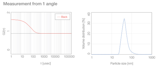
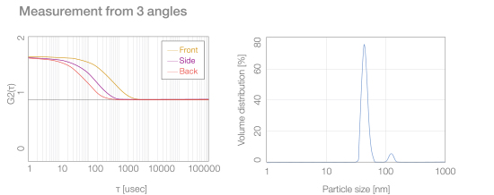
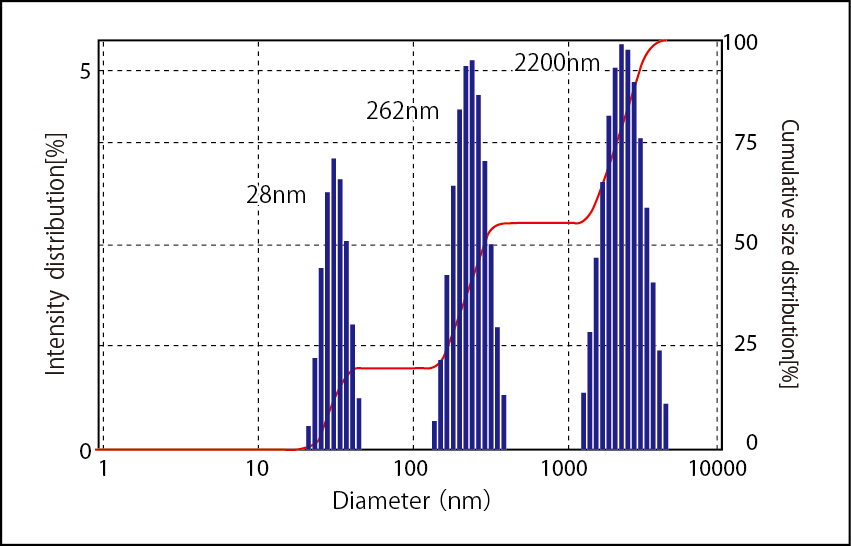
Measurement and analysis from 3 angles - front, side, and back – provides a higher-resolution particle size distribution with higher separability.
Sample peaks that cannot be differentiated with measurement from 1 angle can be separated into individual peaks via measurement and analysis from 3 angles.
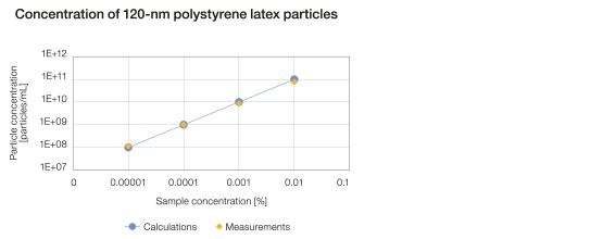
Static light scattering allows calculation of the particle concentration in solution.
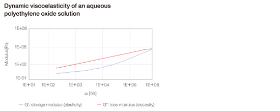
Dynamic light scattering allows measurement of the viscoelasticity of “soft” structures such as polymers and proteins.
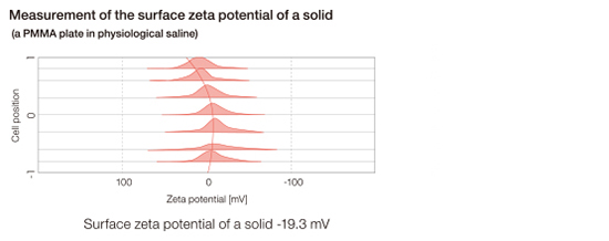
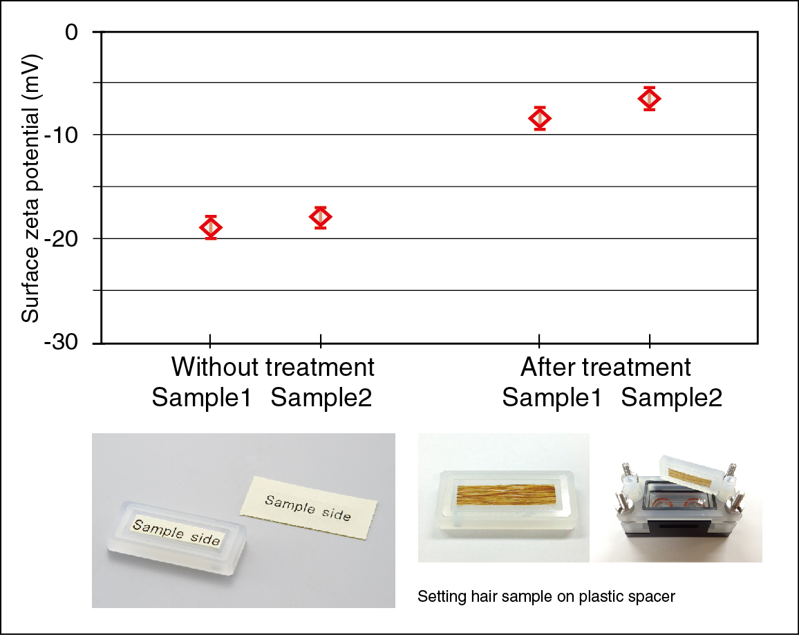
Our flat surface cell has been upgraded to measure the surface zeta potential of flat samples. The cell’s newly developed coating to resist high salt concentrations allows measurement under highly saline conditions (a 154 mM NaCl solution), facilitating the evaluation of biocompatible materials.
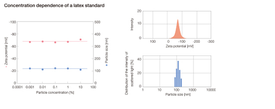
The size and zeta potential of particles can be measured in solutions ranging from a 0.00001% dilute solution (0.1 ppm) to a solution concentrated up to 40%.
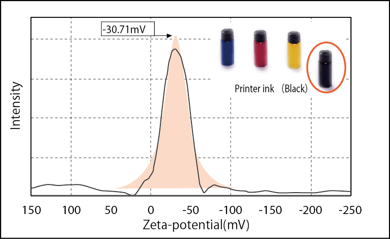
Zeta potential of ink liquid (Black)
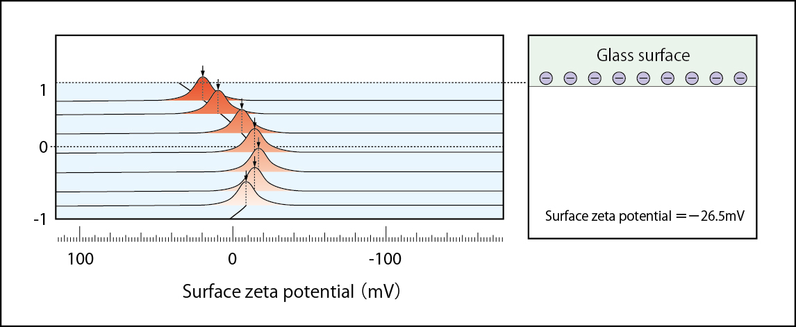
Zeta potential of negatively charged glass surface (BLANK)
Zeta potential =-58.4 mV(1 mM NaCl solution)
Negatively charged glass surface being neutralized by positively charged CTAB
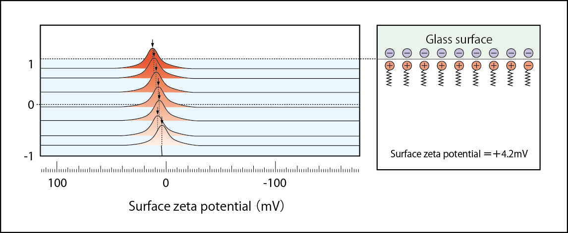
Zeta potential =+1.3 mV(1 mM NaCl solution containing 1×10⁻⁵ mol/l CTAB)
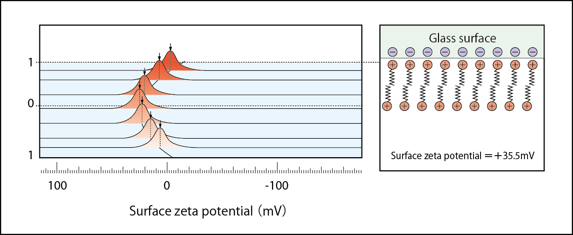
Positively charged status with the excessive CTAB adsorbed on glass surface
Zeta potential =+35.5 mV(1 mM NaCl solution containing 1×10⁻⁴ mol/l CTAB )
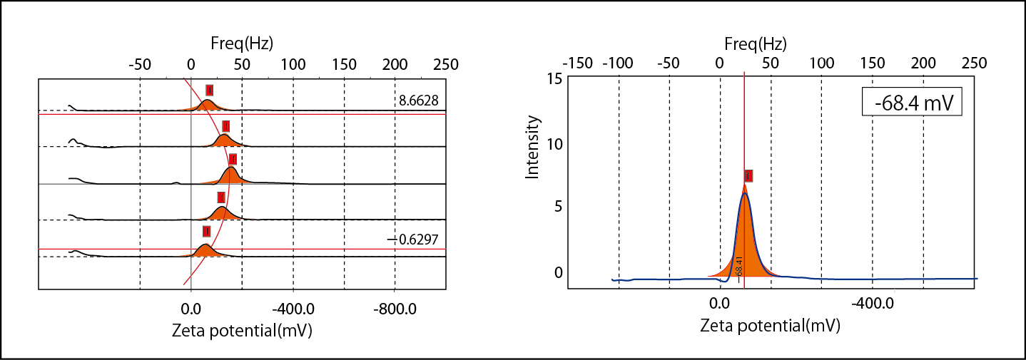
Electro-osmosis plotting and zeta potential of polystyrene latex particles in 100 mM NaCl
Option
Cell units for surface zeta potential measurement of flat samples and films Also for measurement under high salt concentration conditions
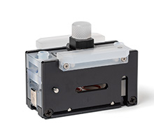
●The cell is easy to assemble
Screw-less design
●A variety of simple coatings
Which Customers can apply coatings by themselves
●Suitable for very small samples
Samples 10×10 mm in size
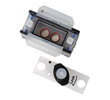
A cell unit for zeta potential measurement with small sample quantities ( 130μL- )
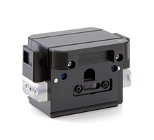
A cell unit for zeta potential measurement in concentrated suspensions
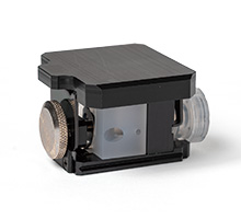
A cell unit for zeta potential measurement in a nonpolar solvent
Suitable for solvents with a dielectric constant lower than 10
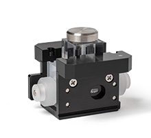
A cell unit for particle size measurement with small sample quantities (3 μL-)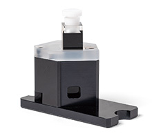
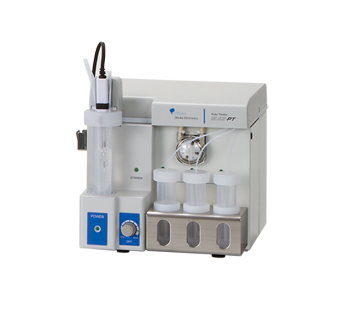
Changes in the size and zeta potential of particles with respect to pH or the concentration of an additive concentration can be measured automatically
- Product
- Principle
- Specification
- Applications
- Option


 Close
Close


